Cell Block a Forgotten Tool
Shaloo Dahima, Padmaraj Hegde, Pushparaja Shetty
Dr. Shaloo Dahima1*, Dr. Padmaraj Hegde2, and Prof. (Dr.) Pushparaja Shetty3
1III MDS, Department of Oral Pathology and Microbiology, A.B. Shetty Dental College, Deralakatte, Mangalore, Karnataka, India
2Assistant Professor, Department of Oral and Maxillofacial Surgery, A.B. Shetty Dental College, Mangalore, Karnataka, India
3Professor and Head, Department of Oral Pathology and Microbiology, A.B. Shetty Dental College, Mangalore, Karnataka, India
- Corresponding Author:
- Dr. Shaloo Dahima
III MDS, Department of Oral Pathology and Microbiology
A.B. Shetty Dental College, Deralakatte, Mangalore
Karnataka, India-575018
E-mail: shaloo.dahima04@gmail.com
Received Date: September 30, 2015; Accepted Date: December 28, 2015; Published Date: December 30, 2015
Citation: Dahima S, Hegde P, Shetty P. Cell Block a Forgotten Tool. J Mol Path Epidemol. 2015, 1:1.
Abstract
Surgical biopsies have always been the mainstay for a precise diagnosis however fine needle aspiration is used as a safe, reliable and an alternative adjunct because of their minimally invasive technique and cost effectiveness. Smears prepared from these aspirates, may at times be inconclusive and difficult to interpret. Hence to make the best possible use of aspirate, smear should be combined with cell block preparation that in turn gives a better morphological and histological detail thus refining the diagnosis. However in the present scenario, cell block technique is still not considered to be a crucial step in the process of histological diagnosis. Here we present case report, where the cell blocks prepared from the aspirate turned out is an important step in diagnosis.
https://marmaris.tours
https://getmarmaristour.com
https://dailytourmarmaris.com
https://marmaristourguide.com
https://marmaris.live
https://marmaris.world
https://marmaris.yachts
Introduction
Swelling in oral and oropharyngeal region is usually a frequent complaint for which patients visit a dental surgeon [1]. The nature of the swellings may vary from being cysts or tumours to inflammatory swellings. Although for precise diagnosis, biopsy remains the mainstay, fine needle aspiration (FNA) has emerged as an adjunct to the this procedure due to its main advantages of being a minimally invasive technique apart from being cost effective, rapid and a safer procedure compared to open surgical biopsy [1]. FNA has been used for diagnosis of palpable masses mainly in breast, lymphnodes, thyroid, prostate, testes and soft tissue tumours and FNA with imaging techniques has been used for diagnosis of non- palpable masses [2]. Despite the advantages of FNA, this technique also presents itself with certain inevitable disadvantages like the material that is aspirated might not be sufficient to make a diagnosis and there is always a risk of false diagnosis or intermediate diagnosis which requires the use of experience and expertise for confident diagnosis using FNA [3]. To make the best possible use of FNA specimen, cell block technique has proved to be of immense use, most important reason being that it is similar to the tissue sections obtained using paraffin blocks thus further clarifying the cellular details and arrive at a diagnosis [3]. Here, we present a case report that asserts the significance of cell block technique in cytopathological diagnosis.
Case Report
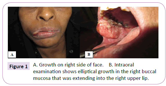
A 45 year old female reported to the Department of Oral and Maxillofacial surgery in January 2015 with a complaint of growth on the right side of the face since 1 month with a gradual progression and history of weight loss. Intraoral examination revealed a 2cm by 3cm elliptical growth in the right buccal mucosa that was extending into the right upper lip. The growth was erythematous, firm and showed everted margins. Tenderness and induration was present (Figure 1). There was no discharge associated with the growth. Ipsilateral Level Ib node was fixed, hard and tender. Patient gave a history of areca nut chewing since 20 years for 3 times a day.
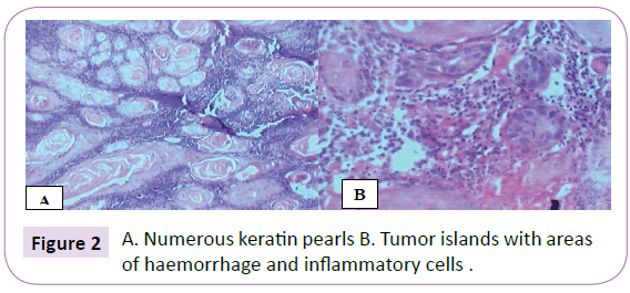
Histopathological examination revealed dysplastic epithelium and connective tissue. Epithelium was stratified squamous parakeratinised of variable thickness and was seen invading the connective tissue in the form of sheets and islands. Epithelial cells showed features of dysplasia like cellular and nuclear pleomorphism, altered nucleocytoplasmic ratio and nuclear hyperchromatism. Numerous keratin pearls were evident (Figure 2). Connective was composed of dense chronic inflammatory cell infiltrate mainly comprising of lymphocytes and plasma cells, adipocytes, blood vessels and extravsated RBCs.
Diagnosis of squamous cell carcinoma was given. A wide surgical excision was done.
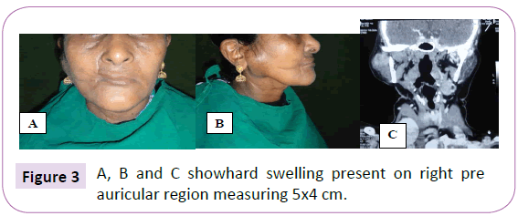
In June 2015, patient again visited Oral and Maxillofacial Surgery Department with a swelling in the right cheek region that was present since 1 month as was reported by the patient. Extraoral examination revealed a hard swelling present on right pre auricular region measuring 5x4 cm which had been increasing in size since the patient first noticed with no pus or blood discharge and no pain present. The mouth opening was adequate however there was deviation of the mandible on the left side while opening the mouth. No intraoral lesion was noticed on examination. CT shows enlarged parotid with multiples heterogeneously enhancing nodules within and multiple subcentric lymph nodes in bilateral level II and left Ib suggestive of intra parotid nodal recurrence (Figure 3).
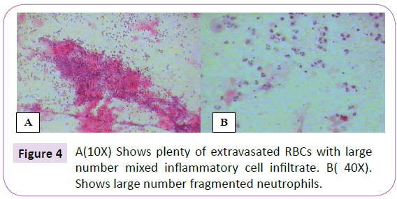
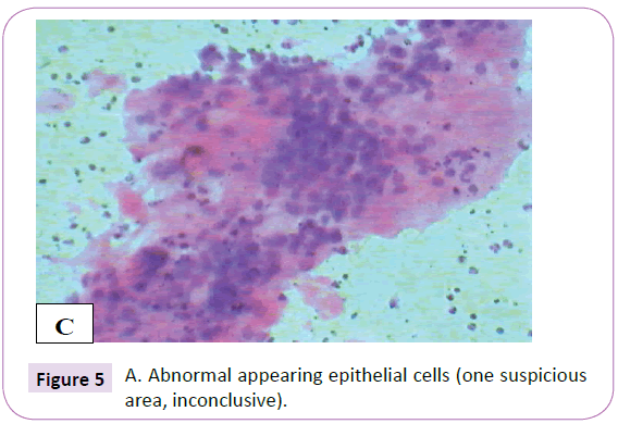
Aspirate was taken from the site and sent to Department of Oral Pathology and Microbiology. Two smears were prepared from the aspirate one for H and E and other for PAP staining. H and E staining revealed plenty of extravasated RBCs with large number mixed inflammatory cell infiltrate in which the majority were fragmented and intact neutrophils (Figure 4). One suspicious area was also present where cluster of cells giving an appearance of abnormal epithelial cells could be seen (Figure 5). PAP smear also showed only the inflammatory cells and extravasated. Hence, these findings proved to be inconclusive.
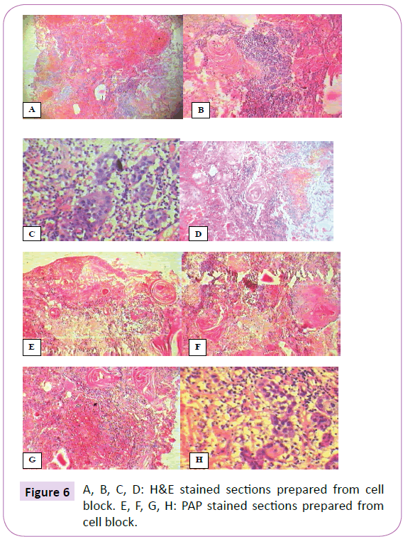
The remaining aspirate was centrifuged and cell block was prepared. The prepared cell block was sectioned in a routine manner followed by staining of the sections by Haematoxylin and Eosin stain (H & E) and PAP or Papanicolaou stain. The cytology by H & E and PAP stain showed similar and highly conclusive picture that was composed of numerous keratin pearls, dysplastic epithelial cells, chronic inflammatory cell infiltrate mainly comprising of lymphocytes and plasma cells, plenty of extravasated RBCs (Figure 6).
Hence, a diagnosis of recurrent squamous cell carcinoma was given and surgical excision was done.
Discussion
For the purpose of cytological diagnosis through an aspirate obtained by the means of fine needle aspiration cytology (FNAC) is considered to be the most reliable, safe and easy to perform. However, its diagnostic efficiency is surpassed by its failure rate that has been mentioned by many to be as high as 45% [4]. The success of fine needle aspiration cytology depends on several technical factors determine its success rate eg. the method of spreading, the thickness of the smear, artifacts due to drying of the smear due to drying in air etc [5-7].
Bahrenburg was the first to introduce cell block technique almost a century ago where he used this technique as an alternative to preparation of conventional smear from ascetic fluid [8]. Since then the cell block has been prepared from various specimens like urine, pleural, peritoneal, ascetic and pericardial fluid as well as from tissue scrapings and hemorrhagic aspirates [9,10]. Cell block technique combines the advantages of fine needle aspiration cytology in being fast and safe method and providing with a precise diagnostic architecture similar to a histological diagnosis [11].
Cell block technique presents with some advantages and disadvantages. Advantages being that the histological pattern of the diseases can be appreciated which is not usually seen in smear preparations and multiple sections can be taken from the cell block that can be subjected to routine or special staining and IHC. During the microscopic examination as well cell block technique poses less difficulty for observation as lesser dispersal of the cells occurs with this technique. Also the like the conventional slides the sections prepared from the cell block can be stored for a longer period of time for restrospective studies as well [12-16].
In the present case also the advantages of the cell block technique hold true for the fact that when we compare the stained sections obtained from the smear and cell block the difference that helped in diagnosis prior to biopsy was clearly visible in the above pictures. The sections prepared from the cell block clearly showed the numerous areas of keratin pearls and areas of dysplastic cells convincing enough to give the diagnosis of squamous cell carcinoma. All these features which were mimicking to those seen in tissue sections were not seen in stained smears as seen in the pictures above.
However cell block technique has some shortcomings the most obvious of which is that the technique is time consuming when compared to conventional smear preparation. It is possible that during the process of centrifugation the material may be lost and may prove to less to come to a conclusive diagnosis. In the process of centrifugation sometimes artifacts can occur in the cell block that may be manifested as rosettes or pseudoacini that may be a source of misdiagnosis [17].
There have been reported cases where the cell block technique has proved to be a useful adjunct with the histopathological diagnosis as it has been in the present case. Cell block technique has been used as a method for presumptive diagnosis of cystic lesions of the jaw like inflammatory cysts where the sections prepared from aspirate and subsequently by cell block revealed cholesterol clefts and inflammation and Keratocystic odontogenic tumour revealed plenty of parakeratin thus helping in distinguishing between the inflammatory to the diagnosis [18].
The cell block has found to be superior in diagnosing neoplasms as well providing a high diagnostic accuracy, however, it has been advocated that the application of smears and cell blocks should be combined wherein the diagnostic accuracy may reach up to 90% especially for neoplastic lesions, to correlate with the histology, as many a times diagnosis met by the cytologic criteria and histologic criteria do not correlate and hence to avoid any pitfalls by using either of them alone, cell block and smears can be used together being complementary in nature to reach a diagnosis [19-21].
Cell blocks as already mentioned can be used for immunohistochemistry and their application can be seen in the detection of HPV status in the head and neck squamous cell carcinoma cases where p16 immunohistochemistry is done on cell-blocks and analysis is done [22].
Bony lesions due to their nature present with very scanty aspirates and more of hemorrhagic aspirates. As the analysis of these aspirates can be difficult cell block technique can be standardised and improvised by various processing techniques and hence overcoming the hindering of diagnosing bony lesions well [11].
To permit good results there is lack of any standardised protocol and hence cell block preparation from fine needle aspiration still takes a backseat compared to smear and histopathological examination, despite the fact that in diagnostic cytopathology cell block technique has an immense significance and can prove to be valuable adjuncts towards a more refined cytological diagnosis complementary to smears and histology [23-25].
References
- Hafez NH, Fahim MI (2014) Diagnostic Accuracy And Pitfalls Of Fine Needle Aspiration Cytology And Scrape Cytology In Oral Cavity Lesions. Russian Open Medical Journal 3:1-8.
- Morse A (2002) Diagnostic Cytopathology: Cell Appearances. In: Bancroft J D, Gamble M. Theory and practice of histological techniques. (5th edition), London, Edinburgh, newyork, oxford, Philadelphia, St Louis, Sydney, Toronto. Churchill livingstone: 637-661.
- Basnet S, Talwar OP (2012) Role of cell block preparation in neoplastic lesions.Journal of Pathology of Nepal. 2:272-276.
- Lee KR, Foster RS, Papillo JL (1987) Fine needle aspiration of the breast. Importance of aspirator. Actacytol31:281-284.
- Sanchez N, Selvaghi SM (2006) Utility of Cell Blocks in the diagnosis ofthyroid Aspirates. Diagn. Cytopathol34:89–92.
- Svante R Orell (1992) Manual and atlas of fine needle aspirationcytology(FNAC).Churchill Livingstone, Newyork: 96pp.
- Chen PK (2004) Artifacts of cytology cell block in fine needle aspirationbiopsy of thyroid. DiagnCytopathol31:362-363.
- Bhatia P, Dey P, Uppal R, Shifa R,Srinivasan R, et al. (2008) Cell blocksfrom scraping of cytology smear: Comparison with conventional cell block.ActaCytol 52: 329-333.
- Bodele AK, Parate SN, WadadekarAA, Bobhate SK, Munshi MM (2003) Diagnostic utility of cell block preparationin reporting fluid cytology. J Cytol20:133-135.
- Qui L, CrapanzanoJP, Saqi A, VidhunR, Vazquez MF (2008) Cell block alone as an ideal preparatory method for hemorrhagicthyroid nodule aspirates procured withoutonsite cytologists. ActaCytol 52:139-144.
- Khan AJ, Hingway SR, Raut WK (2013)A new cost- effective cell block technique forOptimisation of fine needle aspirates in bone Lesions. International journal of medical and applied sciences 2:324-333.
- Thapar M, Mishra RK, Sharma A, Goyal V, Goyal V (2009) A critical analysis of the cellblock versus smear examination in effusions. J Cytol 26:60-64.
- Dekker A, Bupp PA (1978) Cytology of serous effusions. An investigationinto the usefulness of cell blocks versus smears. Am J ClinPathol70:855-860.
- Leung SW, Bedard YC (1993) A simple mini blocktechnique for cytology. Mod Pathol6:630-632.
- Zito FA, Gadaleta CD, Salvatore C, Filático R, Labriola A, et al. (1995) A modified cell block technique for fine needle aspiration cytology.ActaCytol39:93-99.
- Krogerus LA, Anderson LC (1998) A simple method for the preparation ofparaffin-embedded cell blocks from fine needle aspirates, effusions and brushings. ActaCytol32: 585-587.
- Naylor B Pleural (1991)Peritoneal fluids. In: Bibbo M. Editor ComprehensiveCytopathology. (1st Edn), Philadelphia: WB Sounders: 551-621.
- Rivero ERC, Grando LJ, Menegat F, Claus JDP, Xavier FM (2011) Cell block technique as a complementary methodin the clinical diagnosis of cyst-like lesions of thejaw. J Appl Oral Sci19:269-273.
- Tsai YY, Lu SN, Changchien CS, Wang JH, Lee CM, et al. (2002) Combined cytologic andhistologic diagnosis of liver tumors via one-shot aspiration.Hepatogastroenterology 49:644–647.
- Baloch ZW, Tam D, Langer J, Mandel S, LiVolsi LA,et al. (2000) Ultrasound-guided fine-needle aspiration biopsy of the thyroid: roleof on-site assessment and multiple cytologic preparations. DiagnCytopathol23:425–429.
- Basnet S, Talwar OP (2012) Role of cell block preparation in neoplastic lesions. Journal of Pathology of Nepal 2(4): 272 – 276.
- Lastra RR, Pramick MR, Nakashima MO, Weinstein GS, Montone KT, et al. (2013) Adequacy of fine-needle aspiration specimens for human papillomavirus infection molecular testing in head and neck squamous cell carcinoma. CytoJournal10:21.
- Zito FA, Gadaleta CD, Salvatore C,Filotico R, Labriola A, (1995) A modified cell blocktechnique for fine needle aspirationcytology. ActaCytol 39: 93-99.
- Khan S, Omar T, and Michelow P (2012) Effectiveness of the cell block technique in diagnostic cytopathology. J Cytol 29: 177–182.
- Shivakumarswamy U, Arakeri SU, Karigowdar MH, Yelikar BR (2012) Diagnostic utility of the cell block method versus the conventional smear study in pleural fluid cytology. J Cytol29: 11–15.
Open Access Journals
- Aquaculture & Veterinary Science
- Chemistry & Chemical Sciences
- Clinical Sciences
- Engineering
- General Science
- Genetics & Molecular Biology
- Health Care & Nursing
- Immunology & Microbiology
- Materials Science
- Mathematics & Physics
- Medical Sciences
- Neurology & Psychiatry
- Oncology & Cancer Science
- Pharmaceutical Sciences
